NAVIGATE – Fluorescence molecular imaging with rizankizumab-800CW in IBD

Risankizumab is a biologic treatment targeting interleukin 23 (IL-23), used for moderate to severe inflammatory bowel disease (IBD). While effective, predicting individual patient responses remains challenging. This study explores using fluorescently labeled risankizumab-800CW for quantitative fluorescence endoscopy to visualize local drug distribution and target cells, aiming to improve response prediction in IBD patients.
Background
Inflammatory bowel diseases (IBD), including ulcerative colitis (UC) and Crohn’s disease (CD), are chronic, relapsing inflammatory conditions affecting approximately 2.5 million people in Europe. IBD is linked with significant health challenges, complex treatment regimens, and high healthcare costs. A major contributing factor in IBD pathogenesis is the proinflammatory cytokine IL-23. Risankizumab, an anti-IL-23 monoclonal antibody, has been approved for treating moderate to severe CD and recently for UC by the FDA and the EMA Despite the availability of such therapies, many patients exhibit partial or non-responses, with primary non-response rates reaching 30-60% and loss of response over time at 48-58%. Current technologies do not effectively predict treatment responses or visualize drug distribution in tissue, leading to potential exposure to ineffective treatments and adverse effects. To address these limitations, the University Medical Center Groningen (UMCG) developed risankizumab-800CW, a fluorescently labeled version of the drug, to enable visualization and quantification of drug distribution in diseased tissue using fluorescence molecular imaging (qFMI).
Study design
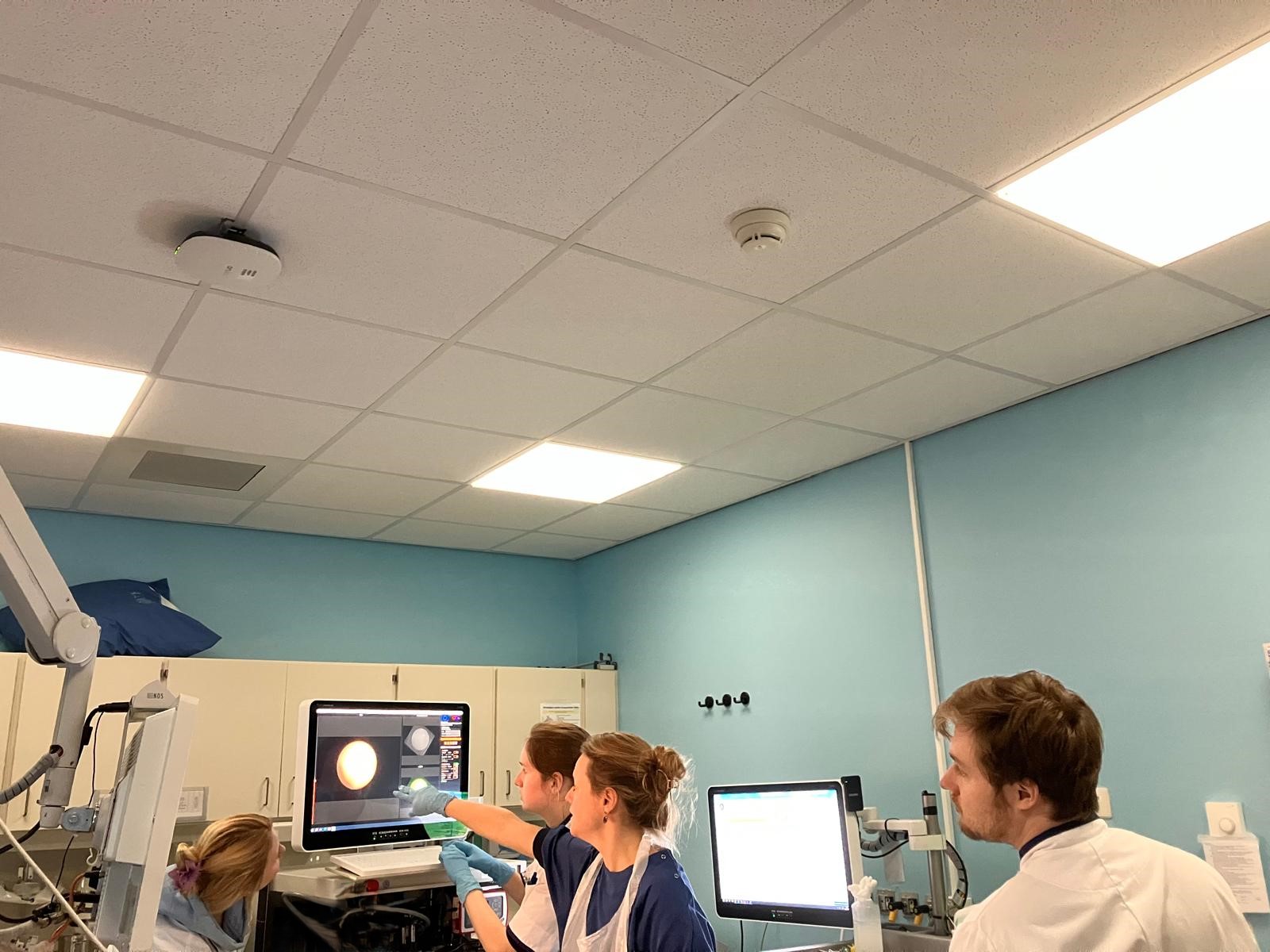
In this study, 18 IBD patients diagnosed with CD or UC and exhibiting active inflammation will be enrolled. Participants will receive an intravenous dose of fluorescently labelled risankizumab-800CW, administered 2–3 days prior to undergoing quantified fluorescence molecular endoscopy (qFME) for real-time visualization of drug distribution. The first part of this study is a dose-escalation phase, starting at 4.5 mg and progressing to 15 mg and 25 mg of risankizumab-800CW, to determine the optimal imaging dose. In the second part patients on risankizumab treatment will get an optimal dose of risankizumab-800CW followed by the qFME procedure. In both parts biopsies will be obtained from both inflamed and non-inflamed mucosal areas for ex vivo analysis, correlating in vivo fluorescence data with histological and molecular findings. Advanced techniques, including spatial transcriptomics, will be used to examine cellular composition and identify risankizumab target cells. Additionally, light-sheet and confocal microscopy, combined with tissue clearing, will facilitate 3D visualization of tracer distribution, providing enhanced insights into drug dynamics within tissues and quantifying the actual tracer concentration.
